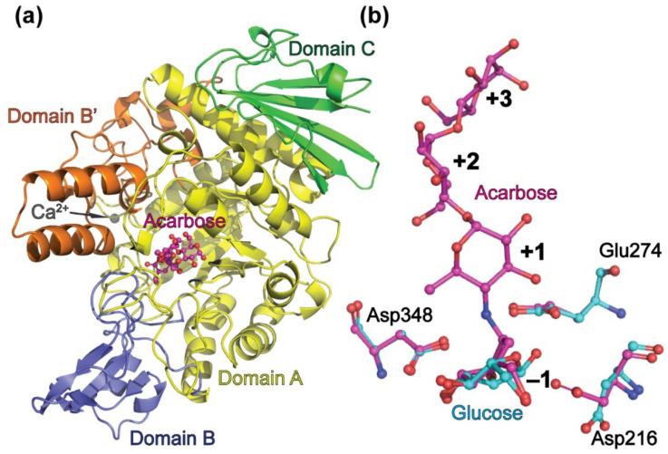
Figure 1.
Overall structure of the BaAG2. A cartoon of the BaAG2-acarbose structure, with acarbose shown in the active center (a). Domain A is in yellow, domains B and B’ are in blue and orange, respectively, and domain C is in green. Calcium ion is shown in grey. Panel (b) shows amino acids of the catalytic triad (Asp216, Glu274, and Asp348) of BaAG2 indicating different orientation of the side chain of Asp216 in the acarbose- and glucose-bound structures. The BaAG2-acarbose structure is shown in magenta, the BaAG2-glucose structure is in light blue. Structures were aligned and visualized with PyMol [39].

