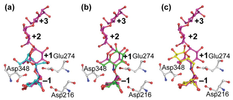
Figure 7.
Positioning of acarbose (magenta) from BaAG2 of B. adeninivorans superimposed with that of sucrose, maltose, and isomaltose. Sucrose (blue, a) originates from the structure of sucrose hydrolase from B. mori (PDB: 6LGF). Maltose (green, b) was taken from the structure of maltase from Bacillus sp. AHU2216 (PDB: 5ZCC) and isomaltose (yellow, c) is from the structure of isomaltase IMA1 of S. cerevisiae (PDB: 3AXH). The catalytic triad of BaAG2 is in grey. Binding subsites of acarbose are labelled with bold numbers. Structures were aligned and visualized with PyMol [39].

