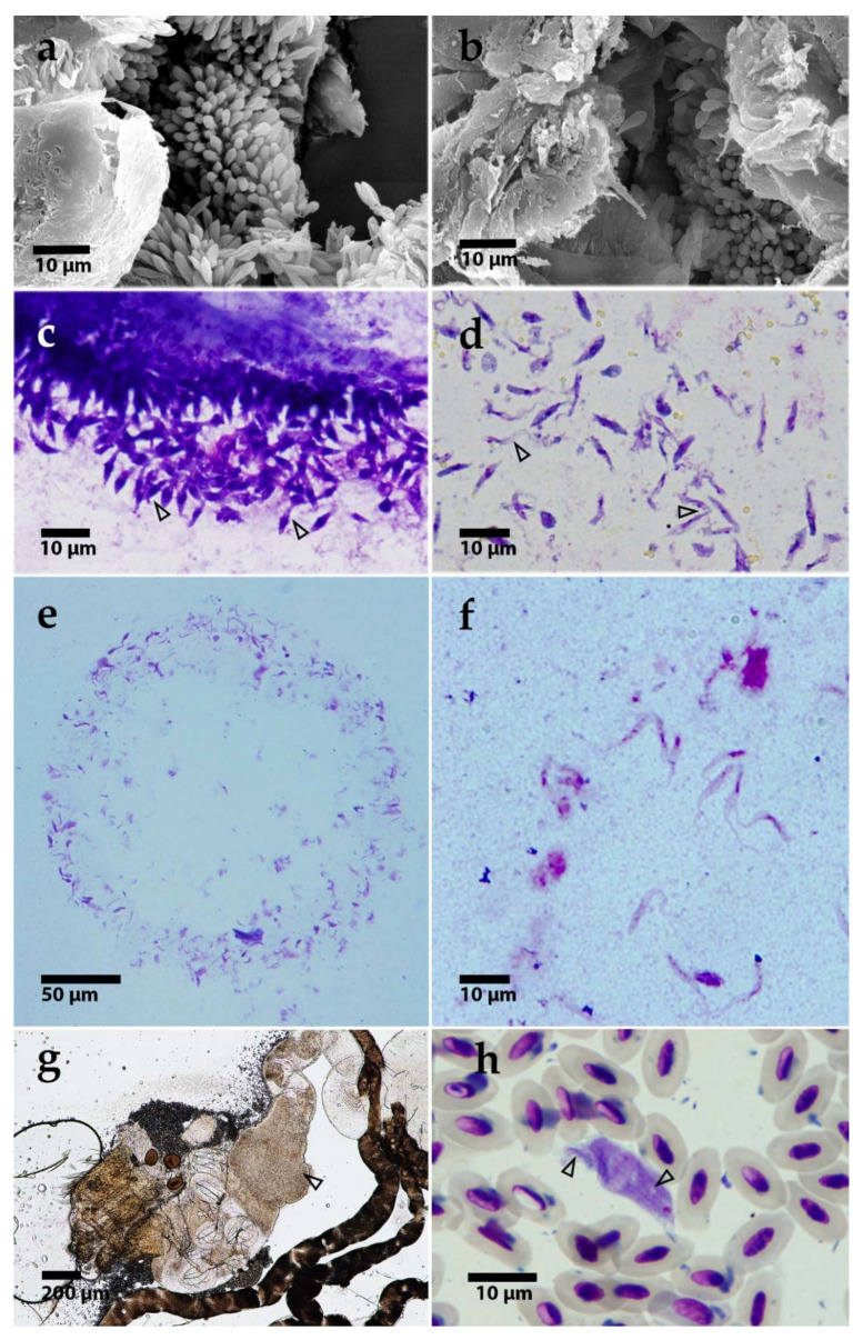
Figure 3.
(a,b) Scanning electron microscopy of T. thomasbancrofti OF19 (a) and PAS343 (b) in the gut of Culex quinquefasciatus after experimental infection (c,d). Light microscopy of trypanosome morphotypes in Culex quinquefasciatus gut: rosettes (c) and epimastigotes (d). Prediuresis droplet with numerous trypanosomes (CUL98) (e). A detail of epimastigotes from prediuresis droplet (f). Dissected Culex mosquito gut with trypanosomes after xenodiagnosis; arrowhead pointing to the mass of parasites (g). T. thomasbancrofti trypomastigote from barn swallow (Hirundo rustica) blood with visible striation (see arrows for striation) (h).

