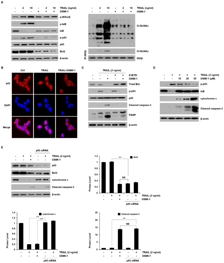
Figure 5.
NF-κB signaling was modulated by TRAIL and OSMI-1 in HCT116. (A) The cells were treated with TRAIL (2 and 10 ng/mL) or combination with OSMI-1 (20 μM) for 24 h. Western blot analysis was performed to detect the levels of p-IKKα/β, p-IκB, IκB, p-p65, p65, Bcl2 (left panel), and O-GlcNAcylated protein (right panel). Total cell lysates were used for immunoprecipitation analysis of IKKβ followed by Western blot analysis with O-GlcNAc antibody (right panel). Equal amounts of the precipitate were used to assess O-GlcNAc levels. (B) HCT116 cells were treated with 2 ng/mL TRAIL for 30 min in the presence or absence of 20 μM OSMI-1. The localization of p65 was visualized using immunofluorescence analysis with anti-p65 (red) and the nucleus was stained with DAPI dye (blue). The merged image represents p65 nuclear transfer. (C) Cells were pretreated with or without Z-IETD-FMK for 1 h and then treated with TRAIL (2 ng/mL) for 24 h in the presence or absence of OSMI-1. Cell lysates were analyzed via western blot analysis using antibodies against Bid, p-p65, p65, cleaved caspase-3 and PARP. (D) Cells were treated with various concentrations of OSMI-1 (10, 20 and 50 μM) in the presence or absence of 2 ng/mL TRAIL for 24 h. Western blot analysis was performed to determine the levels of p-p65, IκB, cytochrome c and cleaved caspase-3. (E) Cells were transfected with control or p65 siRNA and then treated with TRAIL (2 ng/mL) for 24 h in the presence or absence of OSMI-1. The levels of p65, Bcl2, cytochrome c and cleaved caspase-3, were determined via western blot analysis. Data are presented as mean ± SEM, calculated from three biological replicates. *** p< 0.005; NS, not significant upon t-test.

