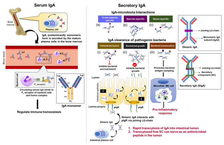
Figure 1.
Structure and functions of serum and secretory IgA. In the left column, IgA, primarily monomeric IgA, is secreted by mature plasma cells in the bone marrow and enters systemic circulation. Circulating serum IgA forms immune complexes with transmembrane Fc receptors located on myeloid cells to induce downstream effector signaling necessary in maintaining immune homeostasis. In the right column, intestinal plasma cells produce dimeric IgA through divalent linkage of two IgA monomers to a joining (J)-chain. The J-chain binds to the secretory component (SC) of polymeric IgA receptors (pIgR) located on the basolateral surface of intestinal epithelial. IgA is rapidly transcytosed into the intestinal lumen as secretory IgA (SIgA). Free SC is also transcytosed into the lumen and serves as an antimicrobial peptide. Interacting with the gut microbiota, SIgA selectivity and reactivity can be categorized into either (a) cross-species (polyreactive) reactive against various bacterial species, (b) species-specific reactivity or (c) strain-specific reactivity. For pathogen removal, SIgA may (i) bind to and agglutinate bacteria, thus hindering microbial attachment and invasion of host intestinal epithelia, a process known as immune exclusion, (ii) prevent bacterial conjugation via enchained growth to limit bacterial proliferation, and (iii) expedite bacterial translocation through microfold (M) cells into Peyer’s patches for antigen sampling by resident dendritic cells (DC).

