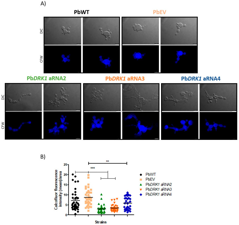
Figure 9.
The level of DRK1 expression reduction correlates with a greater reduction of chitin in P. brasiliensis. Yeast cells of PbWT, PbEV, and PbDRK1 aRNA strains grown for 72 h at 37 °C in BHI medium (1% glucose) (150 rpm). Paraformaldehyde-fixed cells were stained with calcofluor white (50 µg mL−1) for 1 h at RT, and after several washes with PBS were (A) visualized under fluorescence microscopy, DMi8 (Leica) and images processed with LasAF software. Scale bars: 10 µm. (B) The chitin amount was estimated by measuring the calcofluor fluorescence intensity (mean)/area of each cell using ImageJ software. *** p < 0.0001 and ** p < 0.001.

