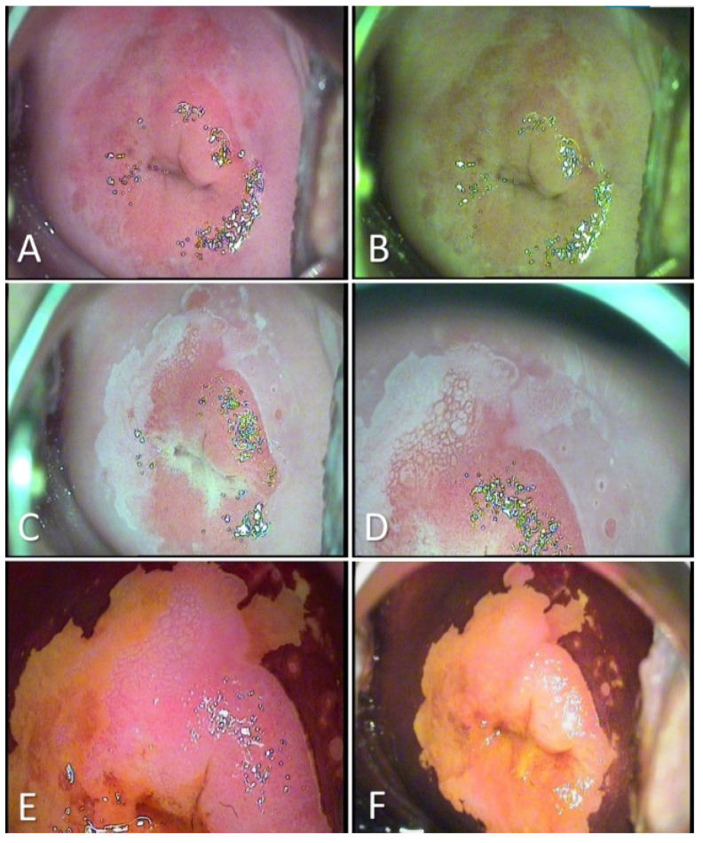
Figure 2.
Colposcopic examination showing HSIL/CIN 2/3 cervical lesions. (A): After the application of saline: Native appearance, no visible lesion; (B): Evaluation in green light filter: does not highlight abnormal vessels; (C,D): After applying acetic acid: Aceto-white epithelium elevated, bright-white, and mosaic visible in three quadrants of the cervix; (E,F): After applying the Lugol solution: iodine-negative lesion, with well-contoured edges and circumferential extension.

