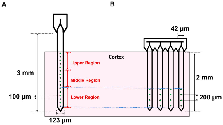
Figure 1.
Dimensions and configurations for single shank and multi-shank microelectrode arrays. Depth-based differences in microelectrode placement for single shank (A) and multi-shank (B) microelectrode devices, highlighting the differences between experimental groups. Pink shaded region indicates the cortex of the brain while dashed lines indicate that microelectrodes in the lower third of single shank arrays are implanted and aligned to the depth of multi-shank microelectrodes.

