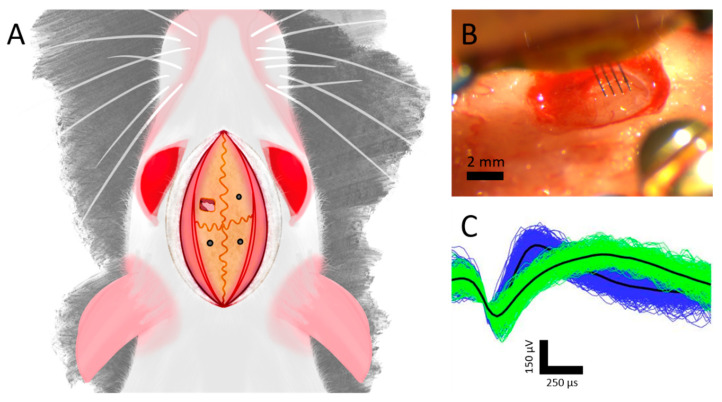
Figure 2.
Neuronal data acquisition and analysis. (A) Implantation schematic with a craniotomy over the left motor cortex and bone screws in the adjacent quadrants. (B) Representative image during a multi-shank implantation. (C) Representative single units recorded simultaneously from the same microelectrode of a single shank device.

