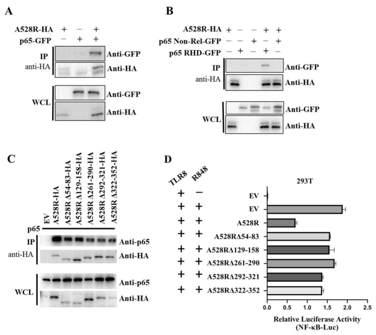
Figure 4.
Interaction between A528R and p65 and analysis of the interaction sites. (A–C) 293T cells in 6-well plate (0.6–1 × 10⁶ cells/well) were transfected A528R-HA (0.5 µg) and p65-GFP (0.5 µg) (A), A528R-HA (0.5 µg) and p65 RHD (0.5 µg) or p65 non-Rel (0.5 µg) (B), A528R ANK deletion mutants (0.5 µg each) (C) for 24 h. The cell lysates were subjected for immunoprecipitation and subsequent Western blotting using the indicated antibodies. (D) 293T cells grown in 96-well plate (3 × 10⁴ cells/well) were transfected by Lipofectamine 2000 with pTLR8 (30 ng), A528R (10 ng), or each A528R ANK deletion mutants (10 ng) plus reporters Fluc (10 ng) and Rluc (0.4 ng), which were normalized to 50 ng/well. At 24 h post transfection, cells were stimulated with R848 (5 µg/mL) for 12 h, followed by the measurement of luciferase activities.

