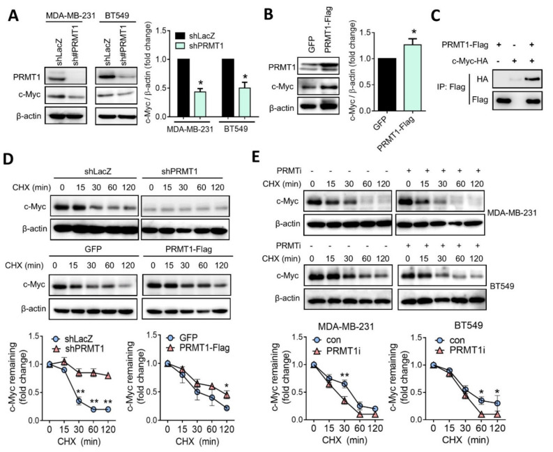
Figure 5.
PRMT1 mediates c-Myc protein level by regulating its stability. (A) Protein levels of c-Myc were analyzed using Western blotting in PRMT1-knockdown MDA-MB-231 and BT549 cells. Relative fold changes of c-Myc to β-actin protein level was shown (right panel). (B) PRMT1 and c-Myc protein levels analyzed using by Western blotting in PRMT1-overexpressing BT549 cells. GFP, green fluorescent protein. BT549 cells were transiently transfected with PRMT1-Flag plasmid, and protein lysates were subjected to Western blot analysis. Relative fold changes of c-Myc to β-actin protein level was shown (right panel). (C) Co-immunoprecipitation analysis of PRMT1-Flag and c-Myc-HA in 293T cells. (D) Western blot analysis of c-Myc protein level in shLacZ and shPRMT1 BT549 cells treated with CHX (50 μM) for the indicated times (0, 15, 30, 60, and 120 min). (Upper panel). Western blot analysis of c-Myc protein levels in GFP and PRMT1-overexpressing BT549 cells treated with CHX (50 μM) for the indicated times (0, 15, 30, 60, and 120 min). (Lower panel). Relative fold changes of c-Myc to β-actin protein levels were shown. (E) c-Myc protein levels were detected using by Western blotting in MDA-MB-231 and BT549 cells following treatment with CHX (50 μM) and PRMT1 inhibitor (40 μM) or CHX (50 μM) alone for the indicated times (0, 15, 30, 60, and 120 min). Western blots were performed from three biological replicates, and quantifications of protein levels were carried out by using Image J software. Unpaired t tests were performed to compare expression levels in two groups. * p < 0.05, ** p < 0.01 were considered statistically significant.

