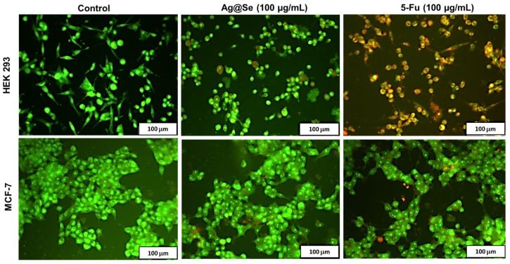
Figure 6.
Fluorescent images of acridine orange/ ethidium bromide dual stained human embryonic kidney (HEK 293) and breast adenocarcinoma (MCF-7) cells. Green fluorescence denotes viable cells, dark red denotes necrotic cells, yellow to orange denotes cells undergoing late apoptosis, while yellow-green nuclei denote early apoptotic cells. Scale bar = 100 µm.

