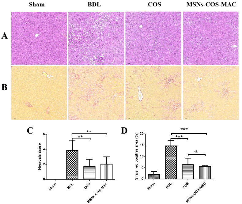
Figure 8.
MSNs-COS-MAC improved liver fibrosis in BDL rats. (A) Liver pathological changes were detected by H&E staining. (B) The degree of liver collagen accumulation was determined by Sirius Red staining. (C) A blinded quantitative assessment hepatic necrosis score. (D) The percentage of Sirius Red positively stained areas. ** p < 0.01 and *** p < 0.001, significantly different from the BDL group.

