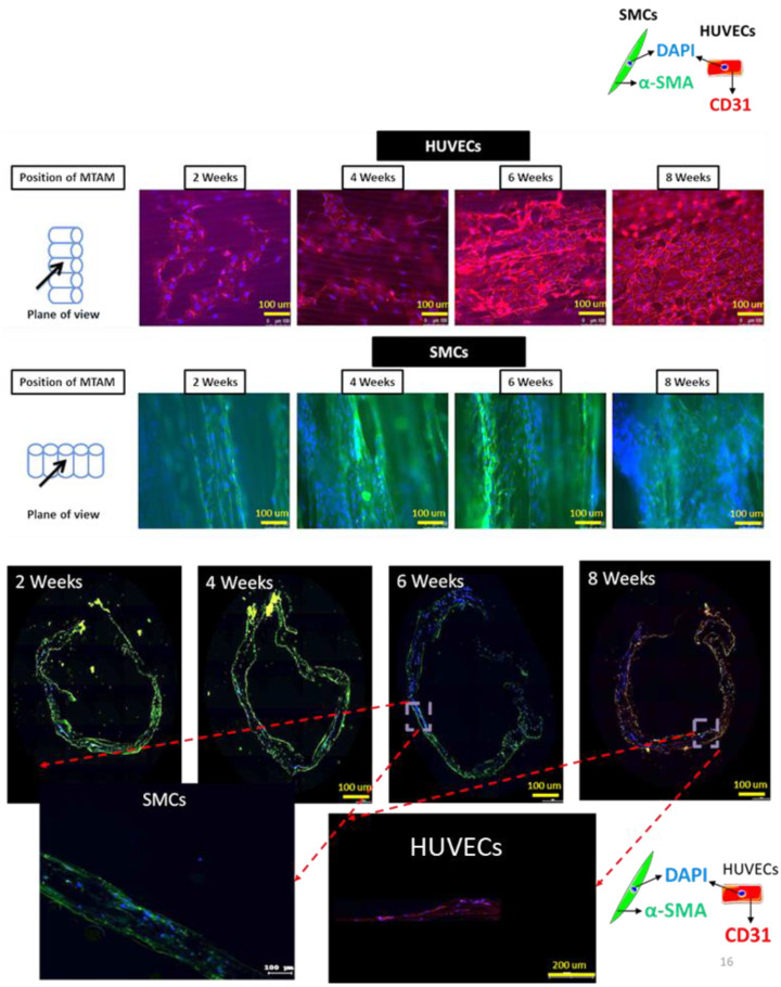
Figure 7.
Fluorescence imaging (FM) of immunocytochemistry stained images of SMCs and HUVECs co-cultured with PLGA MTAMs (top), and fluorescence microscopy imaging of transverse section of 2–8-week old tissue engineered vascular grafts (bottom). A monolayer of HUVECs were observed by week 8 of this study (top, first row), while SMCs proliferated well along the direction of the lumen of the PLGA MTAMs (top, second row).

