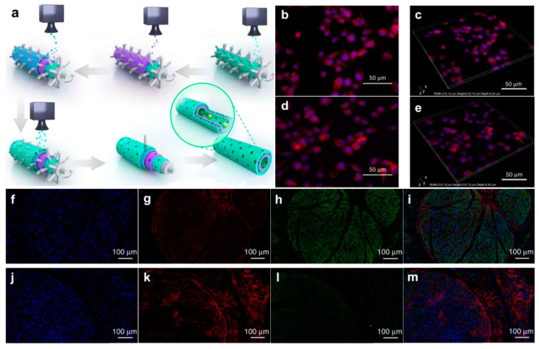
Figure 6.
Fabrication of graphene nerve conduit with LBLC method and its application in axonal regrowth and remyelination. (a) The inner-most and outer-most green layers are PDA/RGD mixed layers. The purple layer is single-layered or multi-layered graphene and PCL mixed layer. The blue layer is a repetition of the graphene and PCL mixed layer. (b–e) Immunofluorescent staining for Ki67 and F-actin. (b,c) Ki67 expression of SC on PDA/RGD-SG/PCL. (d,e) Ki67 expression of SC on PDA/RGD-MG/PCL. (f–m) Triple immunofluorescent staining of Tuj1 and NF200 at 18 weeks post operatively. Tuj1 (green), NF200 (red), and nuclei (blue) were exhibited from different groups, respectively. (f–i) SC-loaded PDA/RGD-SG/PCL. (j–m) SC-loaded PDA/RGD-MG/PCL. Adapted from [55]. Copyright (2018), with permission from Nature Publishing Group.

