Figure 4.
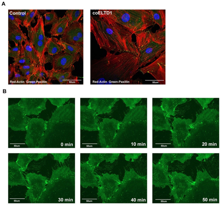
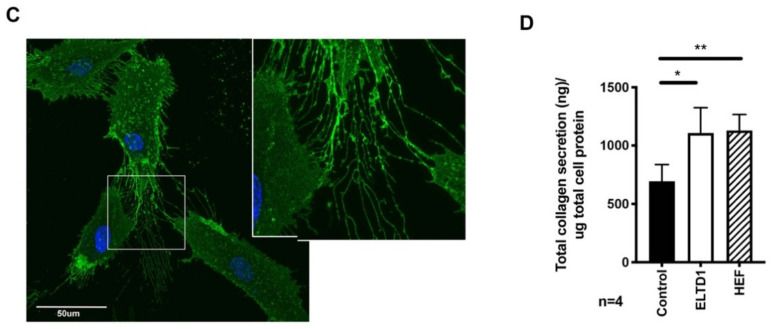
coELTD1 expression induces myofibroblast-like shape changes. (A) Fluorescent staining of actin (phalloidin) and paxillin in control and coELTD1 expressing HUVEC. Images were taken on a Zeiss LSM 880 Confocal Microscope (Zeiss) at 40× magnification. (B) Still images taken from HA-tagged coELTD1 Video S3 at 10 min intervals. HUVEC cells were infected with HA-tagged coELTD1 and imaged 10 min after addition of HA-Alexa Fluor 488 using a Zeiss Observer spinning disc confocal microscope. (C) Membrane staining of non-permeabilized HUVEC cells expressing HA-tagged coELTD1 using anti-HA-Alexa Fluor 488 with zoomed inset of cell tracks. Images were taken on a Zeiss LSM 880 Confocal Microscope (Zeiss) at 63× magnification. (D) Collagen assay performed on supernatant collected from HUVEC cells 120 h after infection with control or coELTD1 virus. * p < 0.05; ** p < 0.005.


