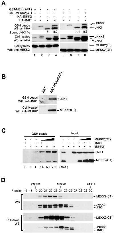
FIG. 4.
MEKK2, JNKK2, and JNK1 form a tripartite complex through synergistic interaction. (A) COS-1 cells were transfected with various combinations of expression vectors for GST-MEKK2(FL) (1 μg/plate) (lanes 1 to 4), GST-MEKK2(CT) (0.5 μg/plate) (lanes 5 to 8), HA-JNKK2 (0.5 μg/plate) (lanes 2, 4, 6, and 8), and HA-JNK1 (0.5 μg/plate) (lanes 3, 4, 7, and 8) as indicated in the figure. GST-MEKK2(FL) and GST-MEKK2(CT) were precipitated with GSH-Sepharose beads, and their associated proteins were analyzed by Western blotting (WB) as described in the legend to Fig. 3. The percentages of HA-JNK1 bound to the GST-MEKK2(CT)-containing complex were measured. Expression of HA-JNKK2 and HA-JNK1 (middle panel) and of GST-MEKK2(FL) and GST-MEKK2(CT) (bottom panel) was determined by Western blotting. (B) COS-1 cells were transfected with GST-MEKK2(CT). After 40 h, GST-MEKK2(CT) was precipitated with GSH-Sepharose beads and its associated proteins were analyzed by Western blotting (WB) using anti-JNK1 antibody. Expression of GST-MEKK2(CT) was determined by Western blotting. (C) Equal amounts of cell lysates prepared from COS-1 cells transfected with GST-JNKK2 vector were mixed with equal amounts of COS-1 cell lysates expressing HA-JNK1 and increasing amounts of HA-MEKK2(CT). The mixtures were incubated for 4 h at 4°C in a rotator, and GST-JNKK2 was precipitated with GSH-Sepharose beads as described above. JNKK2-bound and input HA-JNK1 and HA-MEKK2(CT) were analyzed by Western blotting. The relative fold increase of JNK1 coprecipitated with JNKK2 is indicated. (D) In gel filtration analysis of the MEKK2-JNKK2-JNK1 complex, cell lysates prepared from COS-1 cells transfected with GST-MEKK2(CT), HA-JNKK2, and HA-JNK1 were loaded on a Superdex-200 column and run on a Pharmacia FPLC system. Then 20 μl of each fraction was analyzed by western blotting (WB) with anti-HA antibody and anti-MEKK2 antibody for HA-JNK1, HA-JNKK2, and MEKK2 respectively (top two panels). The remains of each fractions were subjected to a coprecipitation assay with GSH-Sepharose beads as described in the legend to Fig. 3. The precipitated GST-MEKK2(CT) and GST-MEKK2(CT)-bound HA-JNKK2 and HA-JNK1 were analyzed by western blotting (bottom two panels).

