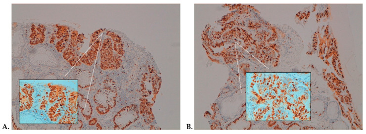
Figure 4.
Immunohistochemistry (IHC) assessment of Ki-67 in high-grade dysplasia showing positive staining in 100% of cells in the pre-treatment sample (A) and 80% of cells post-treatment (B) (magnification: 10× and 40× in the box). Panels A and B are representative of the pre- and post-treatments from the same patient.

