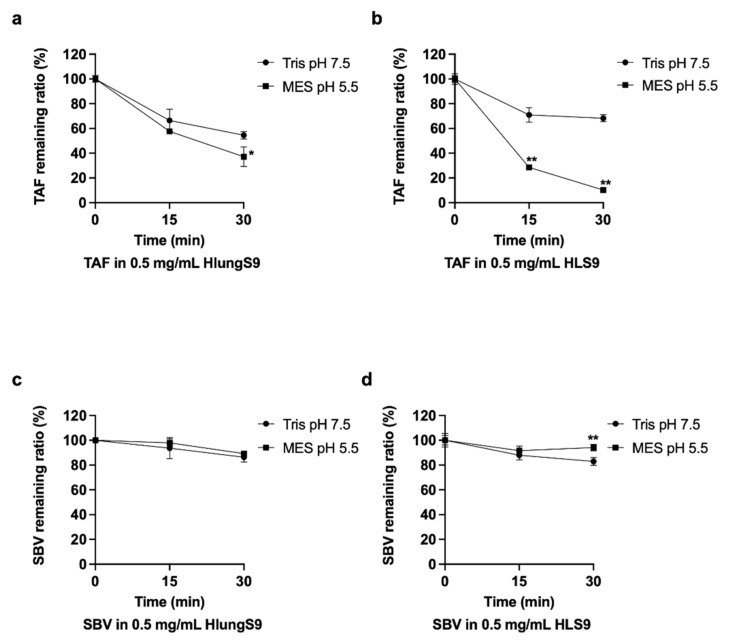
Figure 5.
TAF and SBV hydrolysis in HlungS9 and HLS9: 20 µM TAF were incubated with 0.5 mg/mL HlungS9 (a) and HLS9 (b) at 37 °C for 15 and 30 min in MES assay buffer (pH 5.5) and Tris buffer (pH 7.5); 20 µM SBV were incubated with 0.5 mg/mL HlungS9 (c) and HLS9 (d) at 37 °C for 15 and 30 min in MES assay buffer (pH 5.5) and Tris buffer (pH 7.5). Data are shown as the remaining TAF or SBV (%) after incubations (mean ± S.D., n = 3, except for SBV with 0.5 mg/mL HlungS9 in MES pH 5.5, where n = 2). * p < 0.05 and ** p < 0.01 vs. the Tris pH 7.5 condition (Student’s t-test).

