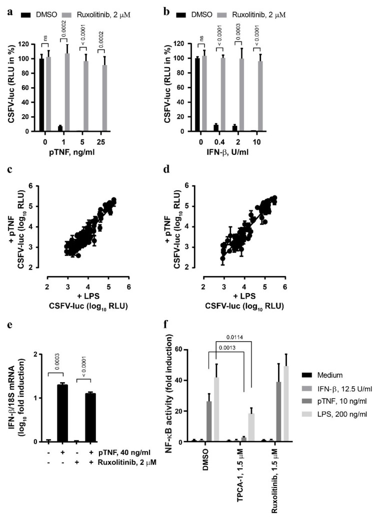
Figure 2.
The anti-CSFV activity of TNF involves JAK/STAT signaling. (a,b) The PEDSV.15 cells were treated with pTNF (a) or IFN-β (b) in the presence of ruxolitinib (2 µM) or DMSO for six hours prior to CSFV-luc infection (MOI, 0.1 TCID50/cell) and measurement of firefly luciferase activity 22 h later. The values are shown as the percentage of the DMSO control in the absence of pTNF or IFN-β, respectively. (c,d) JAK/STAT inhibitor Compound Library screens were performed with CSFV-luc-infected cells pre-stimulated with the medium, LPS or pTNF in the presence of the individual JAK/STAT inhibitors at the concentration of 0.5 µM (c) or 5 µM (d). The data due to cytotoxic and antiviral activity of the compounds were eliminated from the analysis. (e) The PEDSV.15 cells were treated with pTNF or mock in the presence or absence of ruxolitinib for four hours, and the IFN-β mRNA to 18S ribosomal RNA ratio was quantified by means of RT-qPCR and plotted as log10 fold induction. (f) The PEDSV.15 cells were transfected with an NF-κB-dependent firefly luciferase gene reporter plasmid and treated with IFN-β, pTNF, LPS in the presence of the NF-κB signaling inhibitor TPCA-1 or the JAK/STAT inhibitor ruxolitinib. The NF-κB activity is represented as fold luciferase induction related to the medium treatment. The data represent the means and the standard deviations of three independent experimental replicas. The differences were considered to be statistically significant at p < 0.05 using the Student’s t-test (ns, nonsignificant).

