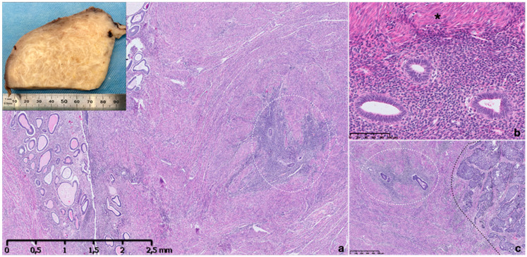
Figure 1.
Macroscopic and microscopic appearance of adenomyosis. Thickened and trabeculated appearing myometrial wall with ill-defined hypertrophic swirls of smooth muscle of sectioned uterus with adenomyosis (a, inset). Histopathological image of uterine adenomyosis observed in hysterectomy specimen, with endometrial glandular and stroma invading the muscular myometrium (within circle) (a). Higher-power view showing ectopic endometrial glands and stroma surrounded by hyperplastic myometrium (asterisk) (b). Ectopic glandular epithelium is proliferative type and stroma is inactive, non-mitotic and composed of monotonous cells (b). A specimen showing an endometroid carcinoma infiltrating myometrial wall on the right (black line) and adenomyosis foci on the left (within line) (c). Hematoxylin and eosin stain.

