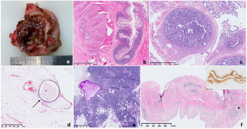
Figure 2.
Macroscopic and microscopic appearance of endometriosis in different sites. Macroscopic picture of ovarian endometrioma with fibrotic wall and a dense, dark brown content (a). Histopathological images of endometriotic implants in intestinal wall, fallopian tube, mesenteric adipose tissue, lymph node and diaphragm (b–f); m: intestinal mucosa; Ft: Fallopian tube; Ly: lymph node. The withe circles and the arrows highlight endometrial implants composed by several endometrial glands and stroma (b–f). Note the presence of hemosiderin-laden macrophages (asterisk) (f). Immunohistochemistry for CD10 shows strong expression in stroma surrounding an ectopic endometrial gland in the diaphragm (f inset). Hematoxylin and eosin stain.

