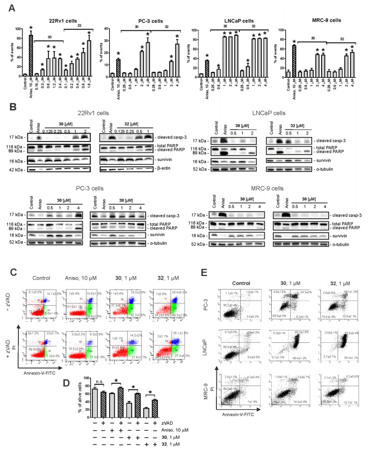
Figure 2.
Pro-apoptotic activity of the synthesized compounds in 22Rv1, PC-3, LNCaP, and MRC-9 cells. (A) Analysis of drug-induced DNA fragmentation using PI staining and flow cytometry. Cells that appeared as sub-G1 population were assumed to be apoptotic and have been quantified with Cell Quest Pro software. (B) Analysis of the expression of pro-apoptotic proteins using Western blotting. β-Actin and α-tubulin were used as loading controls. (C–E) Analysis of apoptotic cells using annexin-V-FITC/PI double staining and flow cytometry. 22Rv1 cells were pre-treated with the pan-caspase inhibitor z-VAD(OMe)-fmk or vehicle (DMSO) for 1 h prior to treatment with the investigated drugs (C,D). PC-3, LNCaP, and MRC-9 cells were treated with the investigated drugs without pre-treatment with z-VAD(OMe)-fmk (E). Cells that appeared in the lower right and upper right quadrants were assumed to be apoptotic. The flow cytometry data were quantified with Cell Quest Pro software; double-negative cells (annexin-V–/PI–) were assumed as alive cells (D). Cells treated with 10 μM anisomycin (Aniso) were used as positive control. In all the experiments, the time of drug exposure was 48 h. Statistical significance: * p < 0.05 (Student’s t-Test, section D; or ANOVA followed by a post hoc Dunnett’s test, section A). n.s.–non significant (p > 0.05).

