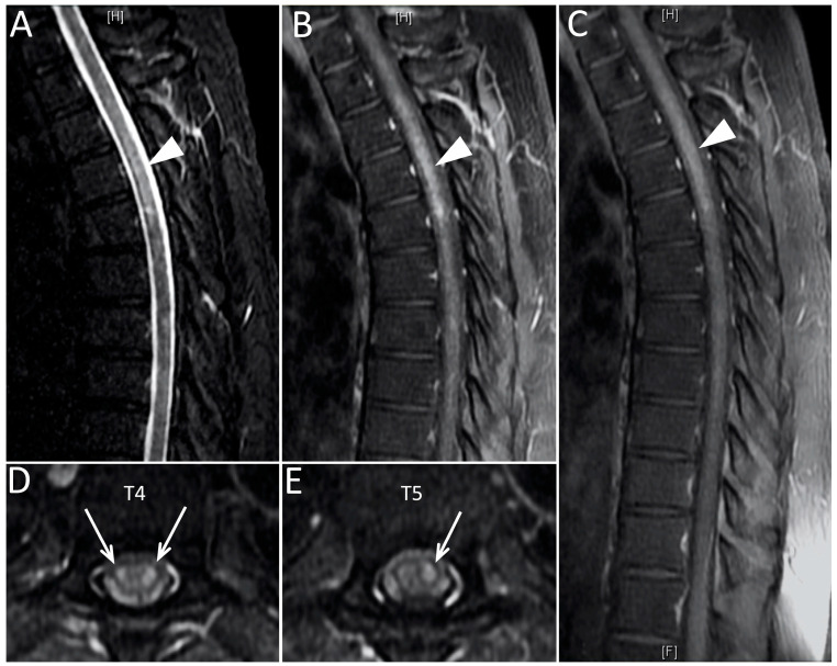
Figure 1.
The MRI of the thoracic spine on the T2-weighted short tau inversion recovery scan (A) showed a hyperintense lesion on T1-T6 (indicated by arrowheads). The lesion was enhanced in the early-phase contrast-enhanced T1-weighted fast spin echo scan (B) and post-contrast T1-weighted fast spin echo scan (C). The axial view of the post-contrast T1-weighted fast spin echo scan over T4 (D) and T5 (E) revealed the enhanced lesion in the spinal cord (indicated by arrows).

