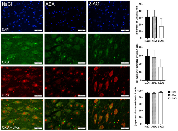
Figure 5.
Immunofluorescence staining and representative images of the lateral hypothalamus (LH) of cows after intraperitoneal administration with NaCl (n = 6), AEA (n = 6) or 2-AG (n = 5) for 9 days. DAPI-stained nuclei (blue), orexin-A neurons (green), cFos (red) and co-localization (merged image). Scale bar within images indicates 20 µm. Number of orexin-A neurons (a), number of cFos-positive orexin-A cells (b) and the percentage of activated orexin-A cells per total orexin-A cells (c). Data are shown as LSM ± SE with (a) F(2,14) = 0.58, p = 0.57, (b) F(2,14) = 0.61, p = 0.56, (c) F(2,13) = 0.45, p = 0.65 (ANOVA).

