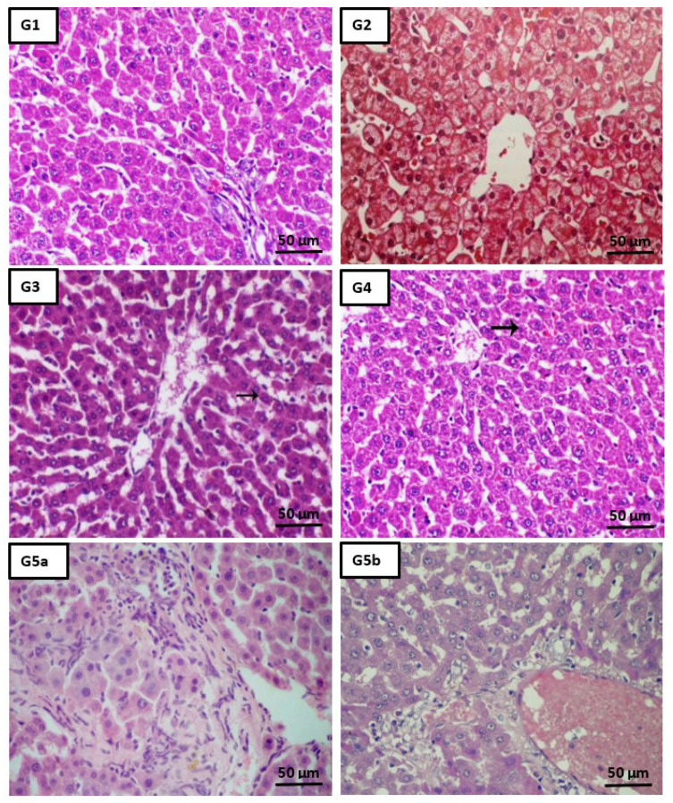
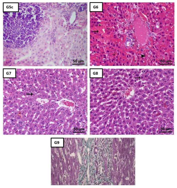
Figure 5.
Effect of different studied compounds on the liver histology. Liver tissues from the control group (G1: H&E, ×200, Scale bar = 50 µm) showed a normal lobular architecture, individual hepatocytes disclosed no pathology, and the portal triad was undetected. Liver tissues of groups CSNPs, DBT, and DBT–CSNPs (G2–G4, respectively: H&E, ×200, Scale bar = 50 µm) exhibited normal hepatocytes around the central vein. A tissue sample from the intoxicated CCl4 group (G5: H&E, ×200, Scale bar = 50 µm) revealed cellular infiltration, congestion of central vein, mild portal inflammation, hemorrhage as well as centrilobular hepatic necrosis, and focus of lytic necrosis & dispersed apoptotic bodies both intra & extracellular in location. The specimen of CSNP-treated rats (G6: H&E, ×200, Scale bar = 50 µm) demonstrated congestion of the central vein and hemorrhage (arrow) and centrilobular hepatic necrosis (arrowhead). The specimen of DBT-treated rats (G7: H&E, ×200, Scale bar = 50 µm) confirmed a mild degree of hepatic degeneration represented by cell swelling (arrow). On the contrary, the liver tissue of DBT–CSNP-treated rats (G8: H&E, ×200, Scale bar = 50 µm) revealed a marked decrease in hepatic degeneration unless single cell degeneration (arrow). Moreover, a periportal inflammatory reaction with a degenerated hepatic cord and disrupted cell plates was observed in the tissue of the cisplatin-treated group (G9: H&E, ×200, Scale bar = 50 µm). These histopathological results revealed the hepatoprotective effects of DBT and DBT–CSNPs, confirming the biochemical analysis.


