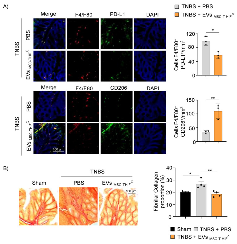
Figure 7.
EVMSC-T-HIFC change the ratio of infiltrating Mφ1/2 and decrease fibrillar collagen content. (A) Immunodetection of F4/F80 (pan-macrophage marker, red) and PD-L1 (pro-inflammatory, Mφ1, green) or CD206 (Mφ2, green) in colon samples 4 days after TNBS-induced colitis. Scale bar: 100 μm. Quantification of double-positive cells per mm2. Ten sections of 0.14 mm2 per mouse were analyzed. Graphs represent the mean ± SEM of three mice. Unpaired t-test was used for statistical analysis. (B) Sirius Red staining was used to detect collagen fibers. Fibrillar collagen content (%) was calculated by dividing red stained area by total tissue area. Scale bar: 100 μm. Graph represents mean ± SEM of four mice. Unpaired t-test was used for statistical analysis. * p < 0.05, ** p < 0.01.

