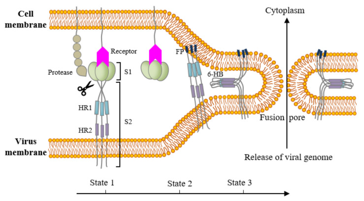
Figure 3.
Schematic model for membrane fusion between coronavirus particles and host cells. State 1 (native): the S1 domain is associated with the S2 domain. The fusion peptide (FP) is buried inside the structure and both of the heptad repeats (HRs) exist as trimers individually. State 2 (intermediate state): the S1 domain is dissociated from the S2 domain after receptor binding. Proteolytic cleavage at the S1/S2 boundary and/or at the S2′ site helps the FP expose and then insert into the cell membrane. State 3 (fusion active state): HR2 folds back to HR1 and forms a stable six-helix bundle, which brings viral and cellular membranes into close proximity, forming a fusion pore, which is followed by releasing viral genome into the cytoplasm of host cells. FP: fusion peptide. HR1: heptad repeat 1. HR2: heptad repeat 2. 6-HB: 6-helix bundle.

