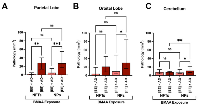
Figure 5.
Alzheimer’s Disease Neuropathology and BMAA Exposure. (A) In the parietal lobe, dolphins with BMAA exposure equivalent or greater than those found in Alzheimer disease (AD) patients ([EE] > AD group, n = 20 tissue sections from 4 dolphins) showed a 14-fold increase in neurofibrillary tangles (NFTs) and a 5.2-fold increase in neuritic plaques (NPs) in comparison to dolphins with less exposure ([EE] < AD; n = 15 tissue sections from 3 dolphins) (**, p = 0.0013; ***, p = 0.0001; ANOVA). (B) The orbital lobe, also showed a 5.5- and 3.2-fold increase in NFTs and NPs, respectively (*, p = 0.0196; ANOVA; ns = no significance). (C) NFT neuropathology in the cerebellum was relatively unchanged with BMAA exposure. However, the density of NPs increased 1.6-fold in the [EE] > AD (**, p = 0.0356; **, p = 0.0052; ns = no significance).

