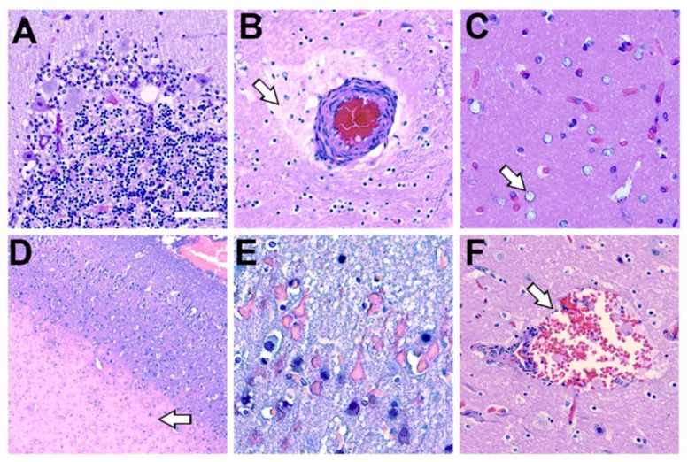
Figure 6.
Histopathology Associated with Methylmercury Toxicity and Dolphin Stranding. (A) Cerebellar Purkinje neurons display disorganization with pallor of the perikaryon (chromatolysis) and vacuolation. In addition, granular cell loss and microcavitation with accompanying gliosis was observed in the cerebellum of dolphin IFAW 12-228 Dd. (B) A blood vessel that displayed thickening of the adventitia and media with expansion of the perivascular spaces, indicative of continuous seepage of serum proteins was observed in IFAW12-228 Dd. (C) Alzheimer’s type-2 astrocytes (arrow), a cellular marker associated with toxin exposure, was observed in the orbital lobe of dolphin IFAW 12-205 Dd. (D) Microvascular lesion in the parietal lobe. The microinfarct has a large area of subcortical necrosis (arrow), with hypoxic-ischemic neurons in the superficial cortical layers of dolphin IFAW 12-198 Dd (E). (F) Subarachnoid hemorrhage observed in the OrL region containing erythrocytes and activated macrophages in Virchow-Robin space (arrow) in dolphin IFAW 12-228 Dd. (40× digital pathology scan; Scale bar = 250 μm).

