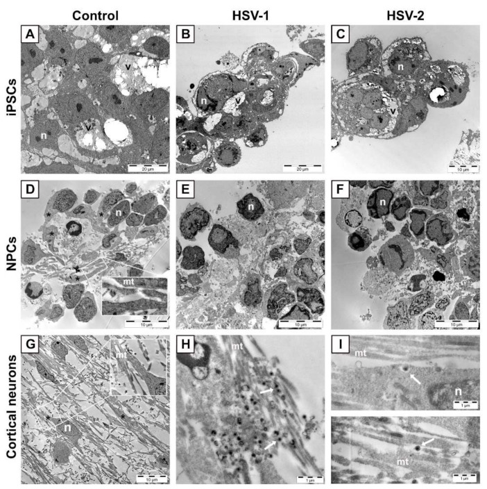
Figure 2.
(A–C) Representative images of iPSCs uninfected or infected with HSV at MOI = 3. (A) Uninfected iPSCs. Large lipid-containing vacuoles (v) are observed in the cytoplasm of iPSCs, independent of infection. Infection of iPSCs with HSV-1 (B) or HSV-2 (C) induces cell rounding 16 h post-infection. (D–F) Representative images of NPCs uninfected or infected with HSV at MOI = 3. (D) Uninfected NPCs show long and wide extensions of the cytoplasm (asterisks) with clearly detectable microtubules (mt in inset) that are lost 24 h post-infection with HSV-1 (E) or HSV-2 (F). (G–I) Representative images of cortical neurons uninfected or infected with HSV at MOI = 3. Neurons display long projections (asterisk) containing microtubules (mt in, inset). These structures are relatively intact 42 h post-infection with HSV-1 (H) and 24 h post-infection with HSV-2 (I). n, nucleus; v, large lipid-containing vacuole.

