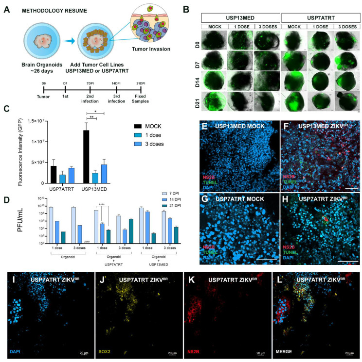
Figure 4.
ZIKVBR infection of CNS tumor cells co-cultured with brain organoids. (A) Schematic representation of brain organoids-CNS tumor cells co-culture assay (DPI: days post-infection). (B,C) fluorescence microscopy analysis of 26-day-old brain organoids co-cultured with 103 GFP+ USP13MED and USP7ATRT cells and treated with none, one or three ZIKVBR doses (B) (scale bar 20 µm). (C) Quantification of GFP fluorescence intensity on 21 DPI images of organoids co-cultured, n = 4. (D) Supernatant PFU assays from co-cultured treated with none, one or three ZIKVBR doses at 7, 14, and 21 DPI. Immunolabeling for non-structural ZIKVBR protein NS2B (red) and cell death (TUNEL, green), and SOX2 (yellow) on 21 DPI images of organoids co-cultured with USP13MED (E,F) and USP7ATRT (G–L), scale bar 50 µm (B,E–H) and 20 µm (I–L). (n = 4 per group) * p ≤ 0.05. ** p ≤ 0.01. **** p ≤ 0.0001.

