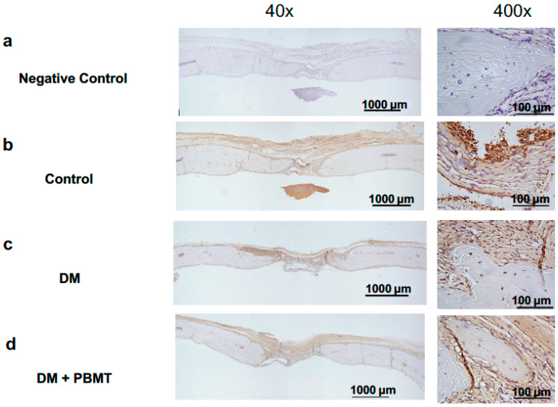
Figure 4.
IHC staining of osteogenic factors BMP-2 of the cross section of the calvarial defects at Week 12 post-operation. Representative BMP-2-stained images of calvarial bone sections from each group: Non-antibody staining control (a), Control (b), DM (c), and DM + PBMT treatment (d). Calvarial bone defects (3 mm) were generated in adult Wistar rats. PBMT treatment (4 J/cm2) was applied daily. The calvarial bone sections were collected at Week 12 postoperatively and stained with BMP-2 antibodies. Scale bar, 1000 μm for 40× and 100 μm for 400×.

