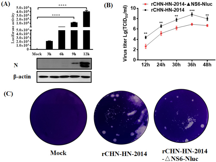
Figure 5.
In vitro growth characterization of the Nluc reporter viruses. (A) Luciferase activity of rCHN-HN-2014-ΔNS6-Nluc-infected cells. LLC-PK1 cells were infected with rCHN-HN-2014-ΔNS6-Nluc, followed by harvesting infected cells at different time point and measuring luciferase activity. Experiments were carried out in triplicate. Collected cell lysates were subjected to Western blot assay using antibodies against N and β-actin. **** p < 0.0001. (B) Multiple-step growth curves of the rCHN-HN-2014 and rCHN-HN-2014-ΔNS6-Nluc on LLC-PK cells. Cells were infected with recombinant PDCoVs at an MOI of 0.01, and cell supernatants were collected at the indicated time points post-infection, followed by TCID50 assays on LLC-PK cells. ** p < 0.01; *** p < 0.001. (C) Mean plaques size of rCHN-HN-2014-ΔNS6-Nluc in LLC-PK cells are smaller compared with that of rCHN-HN-2014.

