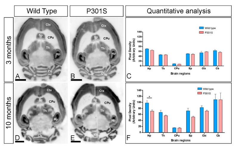
Figure 1.
Regional expression of GIRK2 in the brain of P301S mice. (A–F) The expression of the GIRK2 protein was visualised in histoblots of horizontal brain sections at 3 and 10 months of age in wild type and P301S mice using an affinity-purified anti-GIRK2 antibody. GIRK2 exhibited broad distributions in the brain and region-specific differences were determined by densitometric analysis of the scanned histoblots (panels C,F). Strong GIRK2 staining was observed in the hippocampus (Hp), neocortex (Ctx), cerebellum (Cb), septum (Sp), and thalamus (Th), with moderate staining in midbrain nuclei and faint in the caudate putamen (CPu). Densitometric analysis showed no differences in GIRK2 expression in the brain of P301S mice at 3 months but showed a significant decrease in the hippocampus at 10 months of age (Multiple t-tests and Holm-Sidack method, * p < 0.05). Error bars indicate SEM. Scale bars: 0.2 cm.

