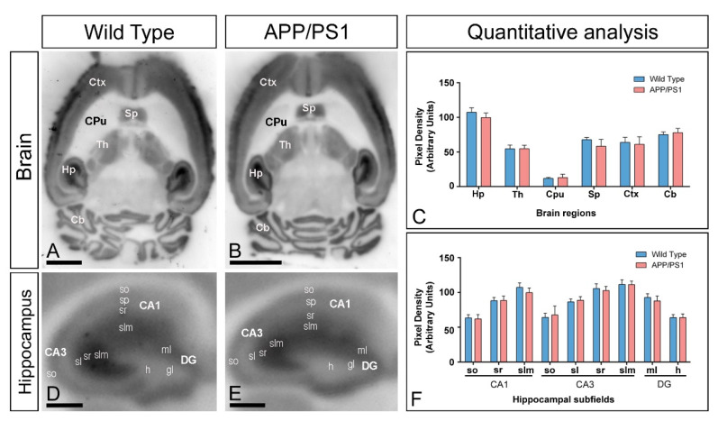
Figure 3.
Regional expression of GIRK2 in the brain of APP/PS1 mice. (A–C) The regional brain expression was visualised in histoblots of horizontal brain sections at 12 months of age in wild type and APP/PS1 mice using an affinity-purified anti-GIRK2 antibody. The expression of GIRK2 revealed marked region-specific differences, with strongest immunoreactivity in the hippocampus (Hp), neocortex (Ctx), cerebellum (Cb), septum (Sp), and thalamus (Th) and weakest in the caudate putamen (CPu). Densitometric analysis showed no differences in GIRK2 expression in APP/PS1 mice compared to age-matched wild type controls. Error bars indicate SEM. (D–F) Hippocampal expression of GIRK2 in wild type and APP/PS1 mice visualised in histoblots of horizontal sections at 12 months of age. Expression for GIRK2 was strong in all dendritic layers of the CA1 and CA3 region and DG, with the strata lacunosum–moleculare (slm) and radiatum (sr) of the CA1 and CA3 regions and molecular layer (mL) of the DG showing the highest expression levels. A moderate expression was observed in the stratum oriens (so) of CA1 and CA3, and the stratum lucidum (sl) of CA3. The hilus (h) of the DG showed the lowest GIRK2 expression level in this region. Densitometric analysis showed no differences in GIRK2 expression in the hippocampal dendritic layers of APP/PS1 mice compared to age-matched wild type controls. Error bars indicate SEM. Abbreviations: CA1 region of the hippocampus; CA3, CA3 region of the hippocampus; DG, dentate gyrus; so, stratum oriens; sp, stratum pyramidale; sr, stratum radiatum; slm, stratum lacunosum-moleculare; ml, molecular layer; gl, granule cell layer; h, hilus. Scale bars: (A,B): 0.2 cm; (D,E): 0.05 cm.

