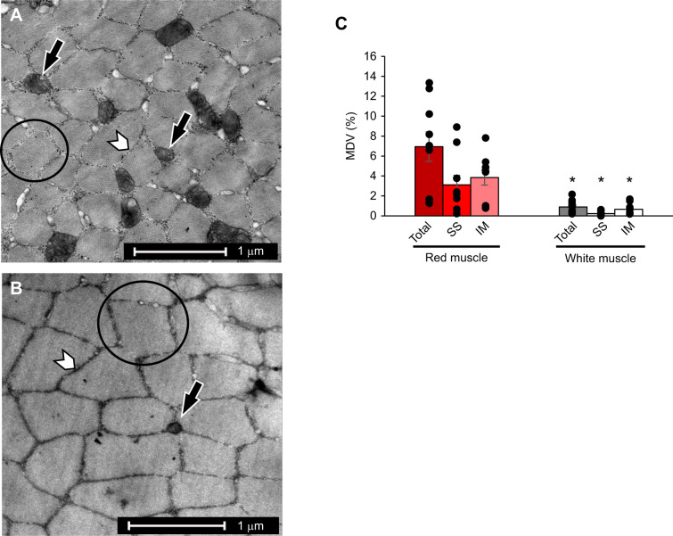
Fig. 1.
Mitochondrial volume density (MVD). Transmission electron micrographs of pinfish aerobic red (A) and anaerobic white (B) skeletal muscle fibers. Black arrows indicate examples of intermyofibrillar (IM) mitochondria, white arrowheads indicate the sarcoplasmic reticulum, and examples of myofibrils are outlined with a circle. (C) Total, subsarcolemmal (SS) and IM MVD in red (n=9) and white (n=11) muscle. Total, SS and IM MVD were significantly greater in red muscle than in white muscle (Student's t-test, *P<0.001, *P=0.006 and *P<0.001, respectively). Data are means±s.e.m.

