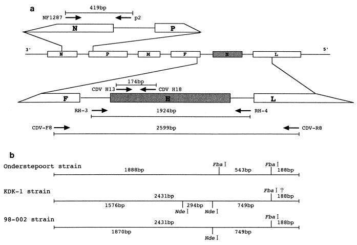
FIG. 1.
Outline of the RT-PCR and scheme for RFLP analysis of the CDV H gene. (a) Genomic organization of the CDV genome, the set of primers, and the lengths of the products of the RT-PCRs. The various CDV genes are represented by boxes. The genes for nucleocapsid protein (N), the phosphoprotein (P), the matrix protein (M), the fusion protein (F), the hemagglutinin protein (H), and the large protein (L) are indicated. (b) Scheme for the restriction enzyme patterns of CDV H genes digested with FbaI and NdeI. Strains Onderstepoort and KDK-1 were used as representative strains of old and new CDVs, respectively. For strain KDK-1, the third restriction site, which was obtained by digestion with NdeI and which generated the smallest fragment (approximately 100 bp), cannot be specified because no sequence information is available for either end of a PCR product for primers CDV-F8 and CDV-R8.

