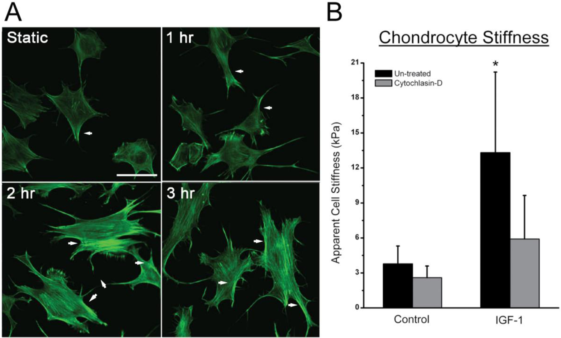
Figure 2: IGF-1 increases actin stress fiber formation and cell stiffness among ATDC5 cells.

A) Time course of IGF-1 treatment indicates an increased stress fiber formation (white arrows) and cell spreading after 1 hour, with the peak amount occurring after 3 hours of treatment. Bar indicates 50 μm. B) Apparent cell stiffness of ATDC5 cells was significantly higher after 3 hours of treatment with IGF-1. The increased stiffness due to IGF-1 was dependent on F-actin organization based on the decrease in cell stiffness following cytochalasin D. (* indicates p-value is < 0.05 compared to controls)
