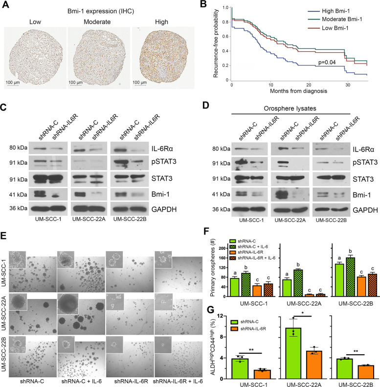
Fig. 1. IL-6/Bmi-1 signaling axis regulates cancer cell self-renewal and correlates with recurrence-free survival of HNSCC patients.
A Immunohistochemistry staining for Bmi-1 in human HNSCC tumor cores of a tissue microarray. Representative images of staining patterns in Bmi-1-low, Bmi-1-moderate, and Bmi-1-high specimens. B Graph depicting adjusted recurrence-free survival function over time in tumors with Bmi-1 expression separated into intensity tertiles. C Western blottings showing baseline protein levels (IL-6R, phosphorylated STAT3, total STAT3, and Bmi-1) in HNSCC cells (UM-SCC-1, -22A, and -22B) stably transduced with lentiviral vectors expressing shRNA-IL-6R or scramble sequence control (shRNA-C). D Western blottings showing protein levels in lysates prepared from primary orospheres generated by HNSCC cells stably transduced with lentiviral vectors expressing shRNA-IL-6R or shRNA-C. E Representative images (×40) of primary orospheres (day 8) formed by IL-6R-silenced or vector control cells. Cells were treated with 20 ng/ml rhIL-6 the day after plating in ultra-low attachment (ULA) orosphere conditions. Inserts at ×100 magnification. F Graph depicting the number of primary orospheres per well. Bar graphs display mean ± SD from five fields per well in three wells per condition. G Flow cytometry graphs depicting the CSC fraction (ALDHhighCD44high cells) in IL-6R knockdown and control cells. Bar graphs display mean ± SD (n = 3). *p < 0.05 or **p < 0.01 as determined by t-test. Different lowercase letters indicate statistical significance at p < 0.05.

