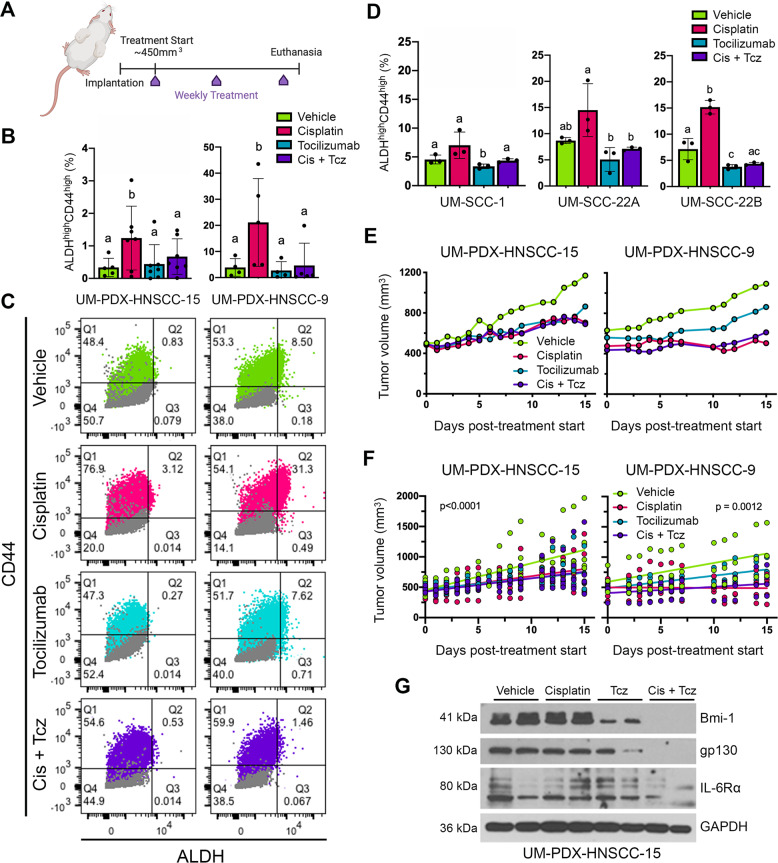
Fig. 2. Tocilizumab suppresses Cisplatin-induced stemness in PDX models of HNSCC in vivo.
A Schematic drawing depicting study design. Mice harboring PDX tumors began weekly treatment for 2 weeks (3 doses total), receiving either no treatment, Cisplatin (5 mg/kg, i.p.), and/or Tocilizumab (10 mg/kg, i.p.). Mice were killed 2 days after last dose. B Bar graphs depicting percentage of CSCs (ALDHhighCD44high cells) in PDX tumors, as measured by flow cytometry. Different lowercase letters indicate statistical significance at p < 0.05. C Representative flow cytometry charts depicting DEAB/IgG controls (gray) for Aldefluor (ALDH activity) and CD44 expression. One experimental replicate per group is shown to demonstrate gate-setting strategy. ALDHhighCD44high cells were identified based on these gates. D Bar graphs depicting the percentage CSCs (ALDHhighCD44high cells) in HNSCC cell lines, as determined by flow cytometry. Bar graphs display mean ± SD (n = 3). Different lowercase letters indicate statistical significance at p < 0.05. E Line graph depicting mean tumor volume over time in the PDX models after treatment with Cisplatin and/or Tocilizumab. Tumor measurements were taken three times per week until study endpoints. F Simple linear regression model of mean tumor volumes over the duration of the experiment. G Western blottings of representative PDX tumor tissue lysates from each treatment group.

