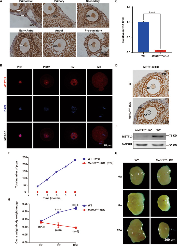
Fig. 1. METTL3 is required for female fertility.
A Immunohistochemistry of METTL3 in 3-week-old mouse ovaries at different follicle stages with PMSG injection. The arrows indicate oocytes at different follicle stages. Scale bar, 20 μm. B Confocal immunofluorescence images of oocytes from wild-type mice stained with METTL3 antibody (red) and DAPI (blue), as indicated. PD5 postnatal days 5, PD12 postnatal days 12, GV germinal vesicle, MII metaphase II. Scale bar, 20 μm. C qRT-PCR analysis of Mettl3 mRNA levels in oocytes from 3-week-old WT and Mettl3Gdf9 cKO females. The relative mRNA level of Mettl3 in WT oocytes was set to 1.0. ***p < 0.001 by two-tailed Student’s t-test. Data represent the mean ± SEM (n = 3). D Immunohistochemistry for METTL3 in ovary sections from WT and Mettl3Gdf9 cKO mice. Primordial (PrF) and secondary (SF) follicle stages are indicated. Scale bar, 20 μm. E Immunoblotting analysis of METTL3 protein level in oocytes of WT and Mettl3Gdf9 cKO mice. GAPDH was used as an internal control. One hundred germinal vesicle oocytes were used for each lane of the blots. F Cumulative numbers of pups born from six pairs of WT and Mettl3Gdf9 cKO female mice for 5 months. G Representative images of ovaries from 6-week-old, 8-week-old, and 12-week-old female mice. Scale bar, 200 μm. H Ratio of ovary weight to body weight of 6-week-old, 8-week-old, and 12-week-old female mice. 6-week-old, n = 3; 8-week-old, n = 6; 12-week-old, n = 6. n.s., p > 0.05; ***p < 0.001 by two-tailed Student’s t-test. Data represent the mean ± SEM.

