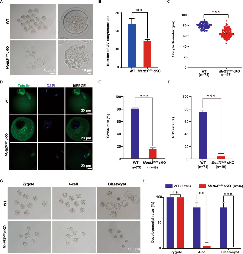
Fig. 3. METTL3 is required for oocyte meiotic maturation and early zygotic development.
A Representative images showing GV stage oocyte from 6-week-old WT and Mettl3Gdf9 cKO females. Left column: scale bar, 100 μm; right column: scale bar, 10 μm. B, C Mean number and diameter of GV stage oocytes obtained per mouse after priming with PMSG. WT, n = 72; Mettl3Gdf9 cKO, n = 97. **p < 0.01; ***p < 0.001 by two-tailed Student’s t-test. Data represent the mean ± SEM. D Confocal immunofluorescence with α-Tubulin antibody (green) and DAPI (blue) for oocytes from 6-week-old WT and Mettl3Gdf9 cKO mice after PMSG and HCG injection. Scale bars, 20 μm. E, F GVBD percentages and first polar body (PB1) rates for oocytes from 6-week-old WT and Mettl3Gdf9 cKO mice after PMSG and HCG injection. WT, n = 73; Mettl3Gdf9 cKO, n = 49. ***p < 0.001 by two-tailed Student’s t-test. Data represent the mean ± SEM. G Representative images of embryos collected from WT and Mettl3Gdf9 cKO mice at the indicated time points after natural ovulation. Scale bar, 100 μm. H Rates of a zygote, four-cell, and blastocyst formation by ovulated WT and Mettl3Gdf9 cKO oocytes after culture in KSOM medium. WT, n = 45; Mettl3Gdf9 cKO, n = 45. n.s., p > 0.05; **p < 0.01; ***p < 0.001 by two-tailed Student’s t-test. Data represent the mean ± SEM.

