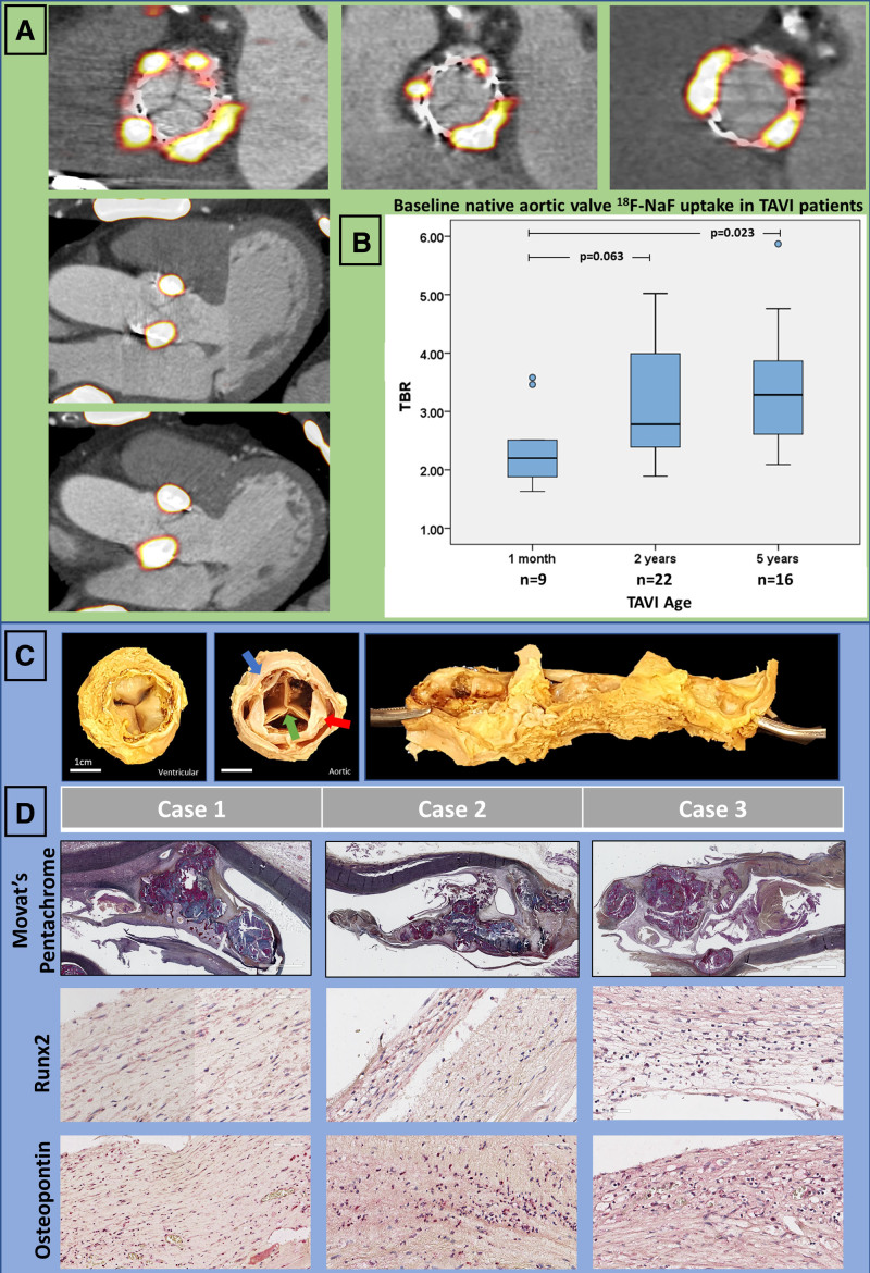
Figure 2.
Baseline assessment with 18F-sodium fluoride activity in native aortic valve tissue after transcatheter aortic valve replacement. A, Hybrid 18F-sodium fluoride (18F-NaF) positron emission tomography and computed tomography en face and long-axis images of native aortic valve tissue uptake. We observed intense tracer activity originating from the native valve tissue around the perimeter of the bioprosthesis in all patients with transcatheter aortic valve implantation (TAVI). B, Native aortic valve 18F-NaF uptake in patients with TAVI was higher with longer duration because bioprosthesis implantation suggesting increased calcification activity after intervention. C, Representative macroscopic images of explanted TAVI valves (green arrow) surrounded by native aortic valve (red arrow) jailed between the bioprostheses and the aortic root (blue arrow): ventricular aspect (Left), aortic aspect (Middle), and view of the root with native valve tissue cut and opened out along its perimeter (Right). D, Histology (Movat pentachrome staining) and immunohistochemistry of native aortic valves showing morphology, high expression of Runx2 and osteopontin in the native aortic valves explanted 1, 32, and 53 months after TAVI. TBR indicates target-to-background ratio.

