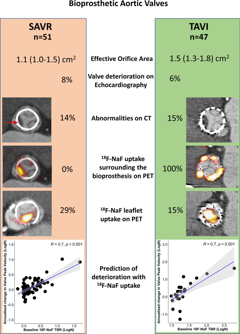
Figure 5.
Comparison of imaging findings and valve deterioration in TAVI vs bioprosthetic SAVR. We compared echocardiographic, computed tomography (CT) and 18F-sodium fluoride (18F-NaF) findings in 47 patients with transcatheter aortic valve implantation (TAVI) with 51 patients with surgical aortic valve replacement (SAVR) who underwent the same research imaging protocol. We observed 18F-NaF uptake on the peripheral of all TAVI valves and none of the SAVR valves. Although patients with TAVI showed lower peak velocity (2.4 [2.0–2.7] vs 2.7 [2.4–3.0] m/s; P=0.03) and larger effective orifice area (1.5 [1.3–1.8] vs 1.1 [1.0–1.5] cm2; P=0.02) than patients with SAVR, we detected baseline echocardiographic (6% vs 8%; P=0.78) and CT abnormalities (15% vs 14%; P=0.87) suggestive of bioprosthetic degeneration in a similar proportion of patients with either TAVI or SAVR. The overall prevalence of patients with increased leaflet 18F-NaF uptake was nearly double in patients with SAVR compared with those with TAVI (29% and 15%; P=0.09). In both patients with SAVR or TAVI, baseline 18F-NaF leaflet uptake was predictive of the change in the peak transvalvular velocity on echocardiography. PET indicates positron emission tomography; and TBR, target-to-background ratio.

