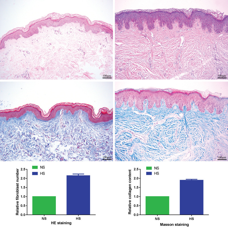
Fig. 1.
Histologic features of hypertrophic scars and normal skin tissues. (Above, left) Hematoxylin and eosin staining for normal skin tissues (original magnification, × 100). (Above, right) Hematoxylin and eosin staining for hypertrophic scar tissues (original magnification, × 100). (Center, left) Masson staining for normal skin tissues (original magnification, × 100). (Center, right) Masson staining for hypertrophic scar tissues (original magnification, × 100). (Below, left) Relative fibroblast number of hematoxylin and eosin staining. (Below, right) Relative collagen content of Masson staining. HS, hypertrophic scar; NS, normal skin; HE, hematoxylin and eosin. Scale bar = 100 μm.

