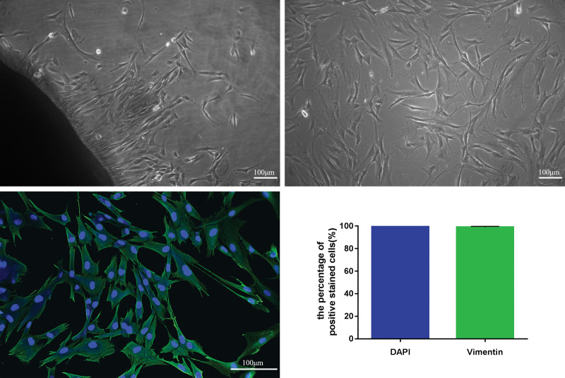
Fig. 3.
Optical and fluorescence micrographs of cell morphologies of hypertrophic scar–derived fibroblasts. (Above, left) The primary fibroblasts were cultured derived from hypertrophic scar tissues (original magnification, × 100). (Above, right) Third-passage fibroblasts derived from hypertrophic scar tissues were observed under inverted microscopy (original magnification, × 100). (Below, left) Cell morphologies of hypertrophic scar–derived fibroblasts were detected by immunofluorescence staining assay (original magnification, × 200). (Below, right) The percentage of positively stained cells. DAPI, 4′,6-diamidino-2-phenylindole. Scale bar = 100 μm (above, right and left, and below, left).

