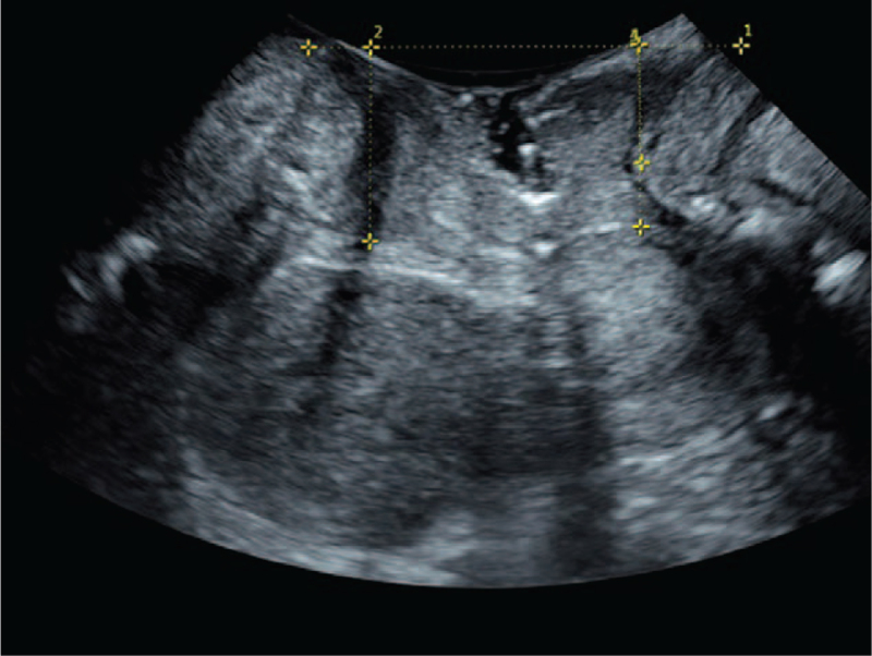
Figure 1.
Ultrasonic image taken in resting state. ∗ Represents that there is a significant difference in the decrease of group B compared with group A (P < .05). Number 1 represents draw a horizontal line through the lower edge of the pubic symphysis in a resting state. Number 2 to 4 represent the distance measurements from the lowest point of the bladder neck (number 2), the lowest edge of the cervix (number 3), and the lowest point of the rectal ampulla (number 4).

