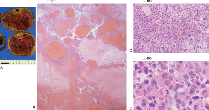
Figure 5.
Histopathological findings of the right adrenal metastasis. (A) Image of the resected specimen obtained after conversion surgery. (B) Hematoxylin and eosin (H&E) staining (×12.5), (C) H&E staining (×100), (D) H&E staining (×400). There was little normal tissue and coagulative necrosis occupied a large area. The tumor cells were slightly more atypical than in the primary tumor.

