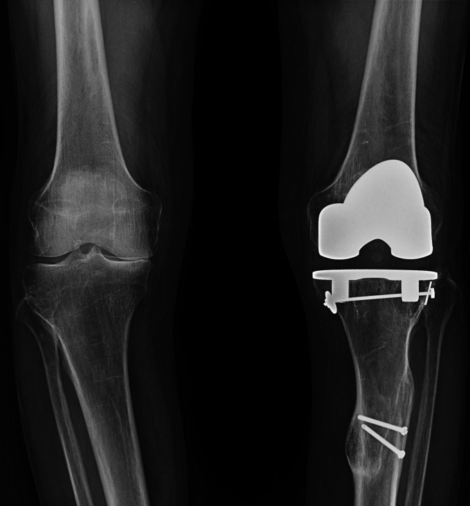
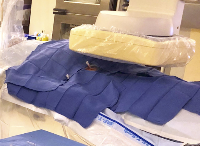
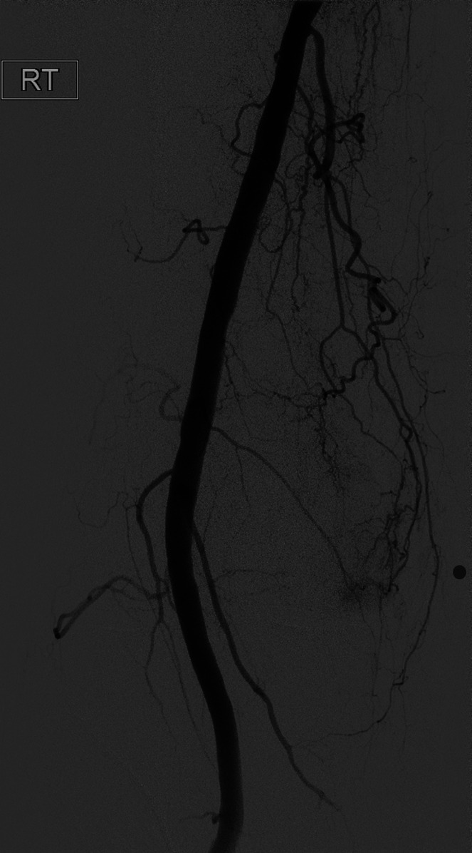
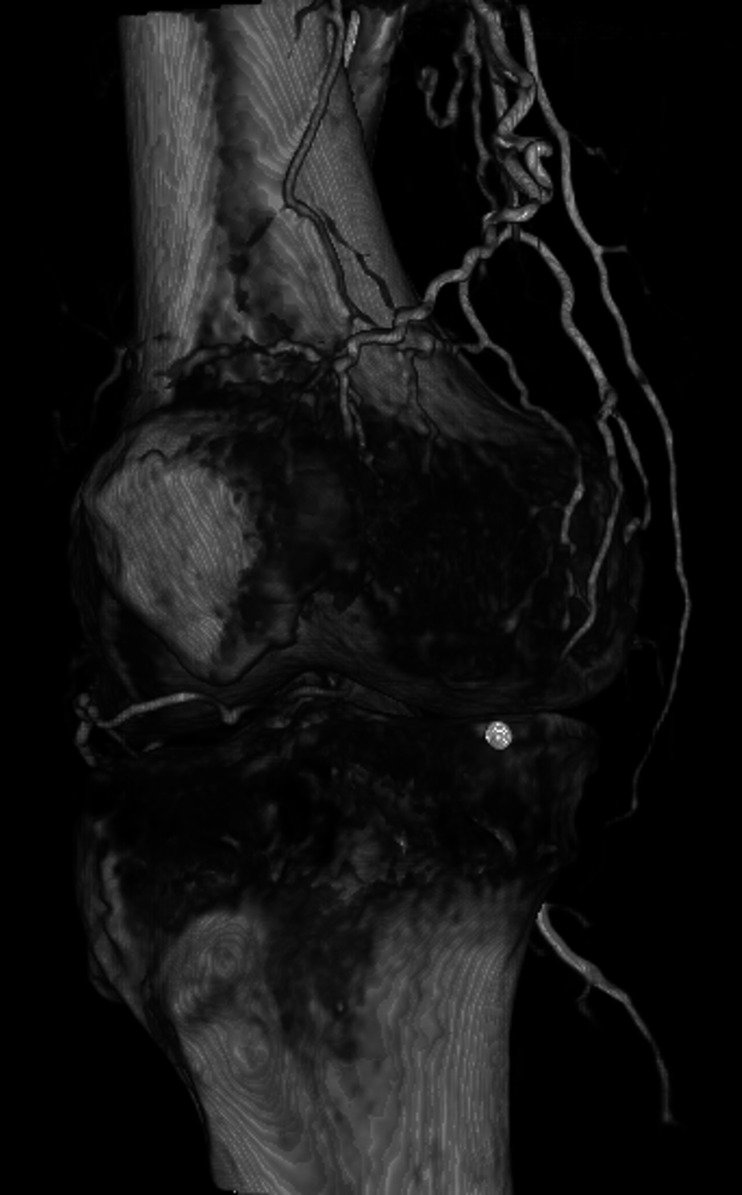
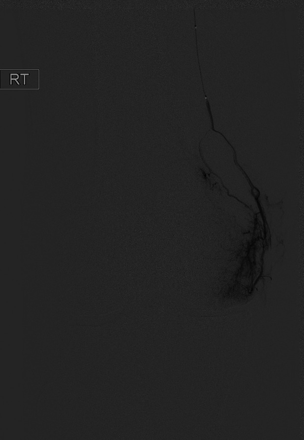
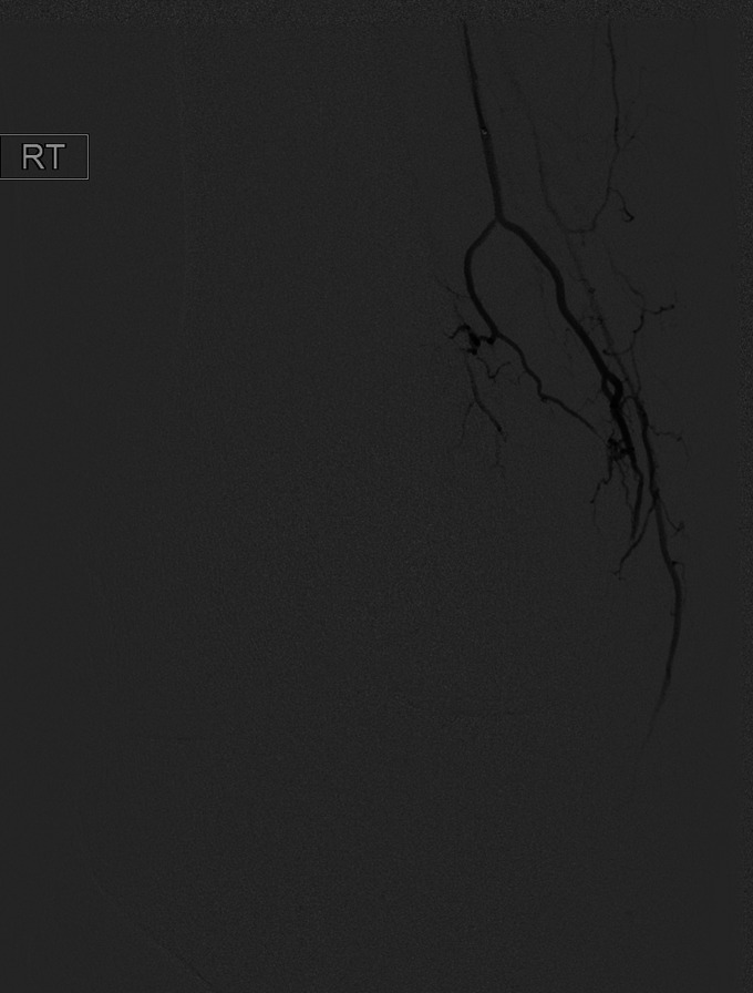
A Figs. 2-A through 2-F A 65-year-old man with pain in the medial aspect of the right knee secondary to OA.
Fig. 2-A.
Fig. 2-A Knee radiograph showing joint-space narrowing of the medial compartment, consistent with Kellgren-Lawrence grade-3 OA.
Fig. 2-B.
Fig. 2-B Access was obtained in the right femoral artery with a 3-French sheath.
Fig. 2-C.
Fig. 2-C Angiogram of the distal superficial femoral artery with a radiopaque marker placed at the site of the pain, showing hypervascularity along the medial joint space.
Fig. 2-D.
Fig. 2-D Rotational 3D reconstructed angiogram identifies the descending genicular artery as coursing toward the region of pain.
Fig. 2-E.
Fig. 2-E Selective catheterization and digital subtraction angiogram of the descending genicular artery confirms the presence of hyperemia in the medial joint space.
Fig. 2-F.
Fig. 2-F After embolization with Embozene microspheres, a postembolization angiogram shows vessel patency with the absence of hyperemia.

